El Paso Times Sept 5 2010
Growing pains: After a long year of excruciating surgeries, 4-foot teen is now a happy 4-foot-6½
Author(s): Victor R. MartinezElsa Gonzalez's arms and legs have too many scars to count. "I know there are plenty of them," she said with a wide grin and a gleam in her eyes. "I wish I had beautiful legs, but this is who I am.
"In a way, I kind of like them. When people stare at them, it gives me the opportunity to tell them what happened to me. I tell them my story, and they are really surprised that I went through what I did."
The 17-year-old junior at Montwood High School has a bone condition known as achondroplasia, a disorder of bone growth that causes the most common type of dwarfism.
In August 2009, Dr. Amr Abdelgawad, an assistant professor of orthopedic surgery and rehabilitation at the Paul L. Foster School of Medicine, put Elsa through four surgeries. The procedure is called limb lengthening, but for Elsa, it was life changing.
El Paso Inc 12/5/2010
Meet El Paso’s new docs
22 percent increase in MDs since 2001
By Robert Gray
Original article published 12/5/2010
Instead of patients having to leave El Paso to get specialized care, the city is beginning to attract patients here to receive the high-tech medicine El Paso’s new doctors are bringing with them.
Doctors like Amr Abdelgawad, 56, whose specialty is pediatric and trauma orthopedics.
Abdelgawad recently helped a 17-year-old junior at Montwood High School grow four inches, even though her body had essentially stopped growing.
Elsa Gonzalez has a condition known as achondroplasia, a disorder of bone growth that causes dwarfism. But after cutting her bone and installing devices used to stretch the bones in her legs, she grew from 50.5 inches to 54.5 inches in one year.
The fixators were removed this March with no complications. Gonzalez could never have had that done in El Paso before Abdelgawad and would have had to travel outside El Paso.
Abdelgawad says he relocated here from Toledo, Ohio, last year after hearing about the city from long-time El Paso orthopedic surgeon Enes Kanlic at an orthopedics conference in Denver.
Abdelgawad visited and decided to stay.
“Having a new medical school and the opportunity to practice trauma and pediatric orthopedics, I thought I could never find something more fitting for me than this,” Abdelgawad says.
He will move into the children’s hospital under construction right now at University Medical Center when it is completed.
El Paso Time Aug 1st 2012:
Juárez boy's surgery part of University Medical Center campaign
By Juan
Antonio Rodriguez / El Paso Times
Posted: 07/31/2012 10:40:20 PM MDT
While most 9-year-old boys run, play and go to school, Eduardo struggles every day to survive.
Eduardo, who lives in Juárez, suffers from severe brain damage, autism, attention-deficit hyperactivity disorder, convulsions and a malformation known as "genu varum," or bowleggedness.
But in the past few months, Eduardo has learned he is not alone in his struggles.
On May 9, Eduardo became the first Mexican boy to have surgery to correct his deformity as part of the University Medical Center Foundation's Borderless Giving Campaign.
The program, started in 2010, focuses on the orthopedic needs of children from Juárez who do not have the economic resources for proper treatment.
"They are children who need help, and they are close to us. I believe we can make a difference in their lives," said Dennece Knight, executive director of the University Medical Center Foundation.
Eduardo, or "Lalito," has lived at the Los Ojos de Dios shelter since 2009. It is at the shelter that he has received individual attention for his neurological and physical needs.
The boy was taken to the shelter because his biological family didn't give him adequate care. Because of his disabilities, his parents made Eduardo live with a cat and a dog, and he shared where they ate and slept.
He was so unattended that he even imitated the animals' behavior.
"Since Eduardo got here three years ago, he has made a huge progress. Now he can eat by himself, walk within his limitations and communicate with others," said Gerardo Rivera, founder of Los Ojos de Dios.
His bowleggedness didn't allow Eduardo to walk normally or by himself.
His left foot was inverted 90 degrees, and as he walked, the other foot also became inverted. He would fall constantly and get hurt.
The Borderless Giving Campaign staff heard about Eduardo through Los Ojos de Dios. The staff helped him get a visa so he could have surgery in El Paso to correct his orthopedic problem.
"We helped to fill out migratory papers and we dealt with medical and legal documents so he could get a visa to get medical treatment," said Julieta Marquez, manager of University Medical Center Foundation in Mexico.
Eduardo's surgery took place May 9 at the El Paso Children's Hospital. Texas Tech pediatric orthopedic surgeon Amr A. Abdelgawad was in charge of the procedure.
"The patient's condition was uncommon," Abdelgawad said. "Generally, the inclination of the feet is only 10 or 20 degrees. We sectioned the bone and we put it in a certain position so he could move it better."
Now three months later, Eduardo has made considerable progress, not only physically but also verbally. He can interact with others.
"He now can rest his feet better. He has more flexibility, and he has even tried to run," said Israel Ortiz, nurse coordinator for Los Ojos de Dios.
Patricia Silis, executive director of Los Ojos de Dios, said Eduardo's surgery was successful and he now will be able to walk better.
Although her institution learned about the University Medical Center program by chance, Silis thinks a superior force makes people meet others at the right moment.
"God is never wrong," Silis said.
Juan Antonio Rodriguez can be reached at Rodriguez@elpasotimes.com.
http://www.healio.com/orthopedics/infection/news/print/orthopedics-today/%7B510E403B-DFFD-4D5C-A7DE-400440BD9CBB%7D/Definitive-early-diagnosis-of-necrotizing-soft-tissue-infections-remains-difficult
Definitive early diagnosis of necrotizing soft tissue infections remains difficult
- Orthopedics Today, September 2012
Although talk of necrotizing soft tissue infections — often referred to as necrotizing fasciitis — is rampant in the media, the incidence rates are low. However, researchers stress that orthopedists should have a high index of suspicion for the condition in every patient with pain or other symptoms that are out of proportion to the initial diagnosis.
See Also
See Also
“I would like all surgeons to be aware of the disease, because you do not diagnose a condition that you do not think about and always have this diagnosis in mind, especially for the elderly and the very young, the people who are intravenous drug abusers or people with hepatitis C infection,”Amr Abdelgawad, MD, an orthopedist with the Paul L. Foster School of Medicine at Texas Tech University Sciences Center in El Paso, told Orthopedics Today. “Have the diagnosis in mind always, and be aggressive.”
Necrotizing fasciitis may be difficult to recognize at presentation because its symptoms often resemble the redness and warmth of synovitis or cellulitis, Abdelgawad said. However, a missed diagnosis could lead to death or amputation for the patient. Russell R. Russo, MD, an orthopedist with Louisiana State University Health Sciences Center, noted the infection can rapidly spread at a rate of 1 cm per hour. With the condition carrying an average mortality rate of 20%, Russo said orthopedists must quickly recognize patients with the condition. Studies show a missed diagnosis of necrotizing fasciitis raises the mortality rate to more than 80%, Abdelgawad said.
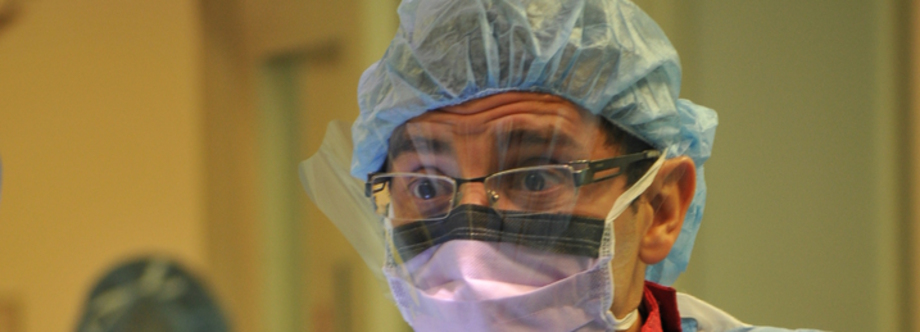
Amr Abdelgawad, MD, said surgeons should keep necrotizing fasciitis in mind when making diagnoses and be aggressive when treating the condition.
Image: Abdelgawad A
“People who have their surgery and debridement within 10 to 24 hours once they hit the door, do much better than the patients for whom the diagnosis is not made for days,” Russo told Orthopedics Today.
Clinical diagnosis, etiology
When patients seem sicker than they appear, orthopedists should have a high index of suspicion,William T. Obremskey, MD, MPH, an orthopedist at Vanderbilt Orthopaedic Institute in Nashville, told Orthopedics Today. A hands-on physical examination is necessary to recognize the condition early, according to Russo, because X-rays, CT scans and other imaging tests may not detect the disease and are time-consuming. Pain out of proportion to physical findings, nausea, fever, hypotension, mental confusion, loss of sensation, skin discoloration, crepitus or necrosis all signal necrotizing fasciitis, Abdelgawad and general surgeon Addison May, MD, FACS, FCCM, of the Division of Trauma and Surgical Care at Vanderbilt University Medical Center, said.
Addison May
When you “put your hand over the extremity, you can feel the crepitus,” Abdelgawad noted.
“Nowadays, people tend to rely on X-rays, CT scans and labs,” he said. “We are getting farther away from going to see patients and putting hands on them — the human touch, the human effect. With [recognizing] necrotizing fasciitis, it is all physical examination.”
Laboratory tests can help detect the condition by determining white blood cell count, erythrocyte sedimentation rate, C-reactive protein, and liver, renal and thyroid function, Russo said. X-rays, CT scans and MRIs may assist orthopedists in detecting gas under the skin associated with the condition, but the tests “are time-consuming and should not delay the patient’s surgical care,” David Hak, MD, MBA, an orthopedist with Denver Health and the University of Colorado, told Orthopedics Today.
“When the diagnosis is suspected, the patient is better treated with immediate surgical exploration rather than further testing,” he said.
Many types of bacteria can cause necrotizing fasciitis, according to Russo and May, including Group A Streptococcus pyogenes and Group BStreptococcus infections. Clostridium and methicillin-resistant Staphylococcus aureus can also cause the illness. In some cases, many types of bacteria can be found in a patient with the condition.
“[One of my patients] had some strep, but she also had some anaerobes, aerobes, gram negative, gram positive — just a host of bacteria,” Russo said.
Predisposing factors
A history of trauma or other predisposing risk factors may help surgeons detect necrotizing fasciitis early, Abdelgawad said. These include obesity, rheumatoid arthritis and immunocompromising conditions such as diabetes, renal disease, HIV, hepatitis C, and bone marrow or organ transplant. However, healthy patients make up 50% of necrotizing fasciitis cases as they can contract the disease from sources such as lacerations, insect bites, endovascular laser treatment for varicose veins or blunt trauma, Abdelgawad said.
“We now see necrotizing fasciitis in healthy kids playing football,” he said. “They are hit in the thigh and all of a sudden have necrotizing fasciitis.”
Obremskey has witnessed the illness in patients with fractures.
“I have seen it in people with closed fractures or in patients with just hematomas under the skin who never had a penetrating injury, but then developed an infection that progressed to necrotizing fasciitis,” he said.
Intravenous drug users are also at higher risk for contracting the disease. Russo’s case of a patient who developed necrotizing fasciitis 2 days after intramuscular injection of the street drug known as bath salts gained national attention after a report in Orthopedics Today’s sister publication Orthopedics. Although the drug is legal in some states, Russo noted that the drug is made in countries where its production may not be regulated. “You do not know if it is in a sterile condition or if the bath salts themselves have bacteria colonized in them,” he said. Some intravenous drug users also “grease needles” — licking the needle prior to injection “which gives you all your bacteria from your mouth, which is one of the most bacteria-loaded places in your body,” Russo said.
However, he warned that used of any improperly sterilized needle — including those used to inject illegal anabolic steroids, epinephrine auto injectors or clean hospital needles — place patients at risk of contracting necrotizing fasciitis.
Surgery and debridement
If a necrotizing infection is suspected, the surgeons agreed that immediate surgical exploration should be performed, with debridement of necrotic tissue and drainage of fluid collections, aggressive resuscitation, and broad spectrum antibiotics to cover the suspected pathogens.
Michael S. Pinzur
Michael S. Pinzur, MD, an orthopedist with Loyola University and Orthopedics Today Editorial Board member, said surgeons should ensure that all of the involved infected tissue is resected. If there is fluid collection, Russo noted that toxins from the infection have already stopped the patient’s blood supply, and antibiotics cannot get through to the infection. In such cases, all the dead vessels, nerves and soft tissues must be surgically removed, he said. Abdelgawad noted that under the skin will mimic “dish water,” because of the foul-smelling discharge of necrotic tissue.
“If you are in doubt, it is much easier to go ahead and open the skin to see if there are any signs of necrotizing fasciitis,” he said. “You would be able to see that the fascia is necrotic, and you are able to move your finger underneath the skin with no resistance.”
Most of the surgeons agreed that one debridement was not enough, and Abdelgawad recommended repeated debridements every 2 days to 3 days as needed. Sometimes, amputation may be necessary. However, if the infection reaches the patient’s neck or abdomen, it is important to seek the help of a general surgeon “because in this case, the mortality rate increases and becomes more than 50%,” Abdelgawad said.
Broad-spectrum antibiotics help prevent super infections, according to May. According to Obremskey, medications that block the immune system’s response to the disease’s exotoxins, such as steroids, can alleviate the illness. Many new immune system blockers are on the horizon, May said, but are 5 years to 10 years away from being introduced to the market.
Wound vacuums, negative pressure wound therapy systems or hyperbaric oxygen may help patients after debridements or amputations, Abdelgawad and Russo said. Intravenous immunoglobulin can be used for cases of sepsis, Russo said, when there are no other options available to attack super antigen helper T cells.
Rehabilitation
Once orthopedists have viable coverage, either by closing the tissue or using skin grafts, they must help patients regain normal function through strengthening exercises, May said. Patient rehabilitation after necrotizing fasciitis must be individualized due to the varying treatments used for the condition including amputation, skin grafts and secondary reconstruction.
“Rehabilitation is based on how much reconstruction you have to do,” Pinzur said. For example, one of Abdelgawad’s patients required an amputation of the whole lower extremity (hip disarticulation) and later on application of a prosthesis, thereby requiring rehabilitation tailored to account for that prosthesis.
In addition, Russo said that patients may require the care of a psychiatrist or other mental health professional for depression and body issues caused by intense pain and limb loss. The dietary departments of hospitals should also be involved and updated on patients’ treatment as nutrition plays a key role in their ability to combat the resultant open wounds and side-effects from antibiotics.
“It is basically a whole body approach to these patients,” Russo said. “They are going to have lots of issues. You need everybody on board to get them up and moving, and get them back to as good as functioning capacity as you can.”
Further studies required
Russo and Abdelgawad agree that researchers must find a way to diagnose necrotizing fasciitis early. In the future, Abdelgawad noted that lab tests or MRIs may enable early diagnosis of the disease.
Russell R. Russo
Russo said a combination of tests to differentiate between cellulitis and necrotizing fasciitis should be developed, and that an immediate ultrasound upon patient admission may help.
“I think if these patients are getting ultrasounds when they hit the door as one of the first tests, you can tell if there is a lot of fluid, pus or gas underneath the skin quickly and rule out cellulitis vs. necrotizing fasciitis relatively quickly without needing a CT, an MRI or a bunch of labs,” he said. “With this disease, time is serious. If you can get an ultrasound in the next room and, running over [the patient’s] affected area you see a pocket of fluid underneath, you can make that diagnosis immediately and get in, get surgery going and significantly reduce patients’ mortality rates and decrease the rates of losing a limb or more.”
May and Obremskey stressed the importance of further immunologic modulation.
“The biggest thing is the interface between our immune system and the bacteria,” May said. “It is that influence of turning our own immune system on and figuring out how to control that,” noting that once the immune system is turned on, it attempts to kill the bacteria, but fights so aggressively that it inadvertently injures the patient’s organs.
Hak and Russo also hold that more education would help new clinicians who never witnessed the disease and patients to recognize it. Russo said 45% of patients do not recall obvious injuries, such as cuts, scrapes, injections or bruises that may have led to infections. He advises telling patients, “If you have a red, warm arm, address it, and do not wait.”
“I think continued [physician] education regarding the condition is still important, since many new clinicians may never see this in their career,” Hak said. “We certainly need better methods to differentiate this condition from the vast majority of less severe soft tissue infections.” – by Renee Blisard Buddle
For more information:
Necrotizing fasciitis may be difficult to recognize at presentation because its symptoms often resemble the redness and warmth of synovitis or cellulitis, Abdelgawad said. However, a missed diagnosis could lead to death or amputation for the patient. Russell R. Russo, MD, an orthopedist with Louisiana State University Health Sciences Center, noted the infection can rapidly spread at a rate of 1 cm per hour. With the condition carrying an average mortality rate of 20%, Russo said orthopedists must quickly recognize patients with the condition. Studies show a missed diagnosis of necrotizing fasciitis raises the mortality rate to more than 80%, Abdelgawad said.
Amr Abdelgawad, MD, said surgeons should keep necrotizing fasciitis in mind when making diagnoses and be aggressive when treating the condition.
Image: Abdelgawad A
“People who have their surgery and debridement within 10 to 24 hours once they hit the door, do much better than the patients for whom the diagnosis is not made for days,” Russo told Orthopedics Today.
Clinical diagnosis, etiology
When patients seem sicker than they appear, orthopedists should have a high index of suspicion,William T. Obremskey, MD, MPH, an orthopedist at Vanderbilt Orthopaedic Institute in Nashville, told Orthopedics Today. A hands-on physical examination is necessary to recognize the condition early, according to Russo, because X-rays, CT scans and other imaging tests may not detect the disease and are time-consuming. Pain out of proportion to physical findings, nausea, fever, hypotension, mental confusion, loss of sensation, skin discoloration, crepitus or necrosis all signal necrotizing fasciitis, Abdelgawad and general surgeon Addison May, MD, FACS, FCCM, of the Division of Trauma and Surgical Care at Vanderbilt University Medical Center, said.
Addison May
When you “put your hand over the extremity, you can feel the crepitus,” Abdelgawad noted.
“Nowadays, people tend to rely on X-rays, CT scans and labs,” he said. “We are getting farther away from going to see patients and putting hands on them — the human touch, the human effect. With [recognizing] necrotizing fasciitis, it is all physical examination.”
Laboratory tests can help detect the condition by determining white blood cell count, erythrocyte sedimentation rate, C-reactive protein, and liver, renal and thyroid function, Russo said. X-rays, CT scans and MRIs may assist orthopedists in detecting gas under the skin associated with the condition, but the tests “are time-consuming and should not delay the patient’s surgical care,” David Hak, MD, MBA, an orthopedist with Denver Health and the University of Colorado, told Orthopedics Today.
“When the diagnosis is suspected, the patient is better treated with immediate surgical exploration rather than further testing,” he said.
Many types of bacteria can cause necrotizing fasciitis, according to Russo and May, including Group A Streptococcus pyogenes and Group BStreptococcus infections. Clostridium and methicillin-resistant Staphylococcus aureus can also cause the illness. In some cases, many types of bacteria can be found in a patient with the condition.
“[One of my patients] had some strep, but she also had some anaerobes, aerobes, gram negative, gram positive — just a host of bacteria,” Russo said.
Predisposing factors
A history of trauma or other predisposing risk factors may help surgeons detect necrotizing fasciitis early, Abdelgawad said. These include obesity, rheumatoid arthritis and immunocompromising conditions such as diabetes, renal disease, HIV, hepatitis C, and bone marrow or organ transplant. However, healthy patients make up 50% of necrotizing fasciitis cases as they can contract the disease from sources such as lacerations, insect bites, endovascular laser treatment for varicose veins or blunt trauma, Abdelgawad said.
“We now see necrotizing fasciitis in healthy kids playing football,” he said. “They are hit in the thigh and all of a sudden have necrotizing fasciitis.”
Obremskey has witnessed the illness in patients with fractures.
“I have seen it in people with closed fractures or in patients with just hematomas under the skin who never had a penetrating injury, but then developed an infection that progressed to necrotizing fasciitis,” he said.
Intravenous drug users are also at higher risk for contracting the disease. Russo’s case of a patient who developed necrotizing fasciitis 2 days after intramuscular injection of the street drug known as bath salts gained national attention after a report in Orthopedics Today’s sister publication Orthopedics. Although the drug is legal in some states, Russo noted that the drug is made in countries where its production may not be regulated. “You do not know if it is in a sterile condition or if the bath salts themselves have bacteria colonized in them,” he said. Some intravenous drug users also “grease needles” — licking the needle prior to injection “which gives you all your bacteria from your mouth, which is one of the most bacteria-loaded places in your body,” Russo said.
However, he warned that used of any improperly sterilized needle — including those used to inject illegal anabolic steroids, epinephrine auto injectors or clean hospital needles — place patients at risk of contracting necrotizing fasciitis.
Surgery and debridement
If a necrotizing infection is suspected, the surgeons agreed that immediate surgical exploration should be performed, with debridement of necrotic tissue and drainage of fluid collections, aggressive resuscitation, and broad spectrum antibiotics to cover the suspected pathogens.
Michael S. Pinzur
Michael S. Pinzur, MD, an orthopedist with Loyola University and Orthopedics Today Editorial Board member, said surgeons should ensure that all of the involved infected tissue is resected. If there is fluid collection, Russo noted that toxins from the infection have already stopped the patient’s blood supply, and antibiotics cannot get through to the infection. In such cases, all the dead vessels, nerves and soft tissues must be surgically removed, he said. Abdelgawad noted that under the skin will mimic “dish water,” because of the foul-smelling discharge of necrotic tissue.
“If you are in doubt, it is much easier to go ahead and open the skin to see if there are any signs of necrotizing fasciitis,” he said. “You would be able to see that the fascia is necrotic, and you are able to move your finger underneath the skin with no resistance.”
Most of the surgeons agreed that one debridement was not enough, and Abdelgawad recommended repeated debridements every 2 days to 3 days as needed. Sometimes, amputation may be necessary. However, if the infection reaches the patient’s neck or abdomen, it is important to seek the help of a general surgeon “because in this case, the mortality rate increases and becomes more than 50%,” Abdelgawad said.
Broad-spectrum antibiotics help prevent super infections, according to May. According to Obremskey, medications that block the immune system’s response to the disease’s exotoxins, such as steroids, can alleviate the illness. Many new immune system blockers are on the horizon, May said, but are 5 years to 10 years away from being introduced to the market.
Wound vacuums, negative pressure wound therapy systems or hyperbaric oxygen may help patients after debridements or amputations, Abdelgawad and Russo said. Intravenous immunoglobulin can be used for cases of sepsis, Russo said, when there are no other options available to attack super antigen helper T cells.
Rehabilitation
Once orthopedists have viable coverage, either by closing the tissue or using skin grafts, they must help patients regain normal function through strengthening exercises, May said. Patient rehabilitation after necrotizing fasciitis must be individualized due to the varying treatments used for the condition including amputation, skin grafts and secondary reconstruction.
“Rehabilitation is based on how much reconstruction you have to do,” Pinzur said. For example, one of Abdelgawad’s patients required an amputation of the whole lower extremity (hip disarticulation) and later on application of a prosthesis, thereby requiring rehabilitation tailored to account for that prosthesis.
In addition, Russo said that patients may require the care of a psychiatrist or other mental health professional for depression and body issues caused by intense pain and limb loss. The dietary departments of hospitals should also be involved and updated on patients’ treatment as nutrition plays a key role in their ability to combat the resultant open wounds and side-effects from antibiotics.
“It is basically a whole body approach to these patients,” Russo said. “They are going to have lots of issues. You need everybody on board to get them up and moving, and get them back to as good as functioning capacity as you can.”
Further studies required
Russo and Abdelgawad agree that researchers must find a way to diagnose necrotizing fasciitis early. In the future, Abdelgawad noted that lab tests or MRIs may enable early diagnosis of the disease.
Russell R. Russo
Russo said a combination of tests to differentiate between cellulitis and necrotizing fasciitis should be developed, and that an immediate ultrasound upon patient admission may help.
“I think if these patients are getting ultrasounds when they hit the door as one of the first tests, you can tell if there is a lot of fluid, pus or gas underneath the skin quickly and rule out cellulitis vs. necrotizing fasciitis relatively quickly without needing a CT, an MRI or a bunch of labs,” he said. “With this disease, time is serious. If you can get an ultrasound in the next room and, running over [the patient’s] affected area you see a pocket of fluid underneath, you can make that diagnosis immediately and get in, get surgery going and significantly reduce patients’ mortality rates and decrease the rates of losing a limb or more.”
May and Obremskey stressed the importance of further immunologic modulation.
“The biggest thing is the interface between our immune system and the bacteria,” May said. “It is that influence of turning our own immune system on and figuring out how to control that,” noting that once the immune system is turned on, it attempts to kill the bacteria, but fights so aggressively that it inadvertently injures the patient’s organs.
Hak and Russo also hold that more education would help new clinicians who never witnessed the disease and patients to recognize it. Russo said 45% of patients do not recall obvious injuries, such as cuts, scrapes, injections or bruises that may have led to infections. He advises telling patients, “If you have a red, warm arm, address it, and do not wait.”
“I think continued [physician] education regarding the condition is still important, since many new clinicians may never see this in their career,” Hak said. “We certainly need better methods to differentiate this condition from the vast majority of less severe soft tissue infections.” – by Renee Blisard Buddle
For more information:
- Amr Abdelgawad, MD, can be reached at the Department of Orthopedic Surgery & Rehabilitation, Paul L. Foster School of Medicine, Texas Tech University Sciences Center, 4801 Alberta Ave., El Paso, TX 79905; email: amr.abdelgawad@ttuhsc.edu.
- David Hak, MD, MBA, can be reached at the Department of Orthopaedics, Denver Health/University of Colorado, 777 Bannock St., MC 0188, Denver, CO 80204; email: david.hak@dhha.org.
- Addison May, MD, FACS, FCCM, Division of Trauma and Surgical Critical Care, Vanderbilt University Medical Center, 1301 Medical Center Dr., Suite 3501, Nashville, TN 37232; email:vicki.moor@vanderbilt.edu.
- William T. Obremskey, MD, MPH, can be reached at Vanderbilt Orthopaedic Institute, 1215 21st Ave. S, Ste. 4200, Nashville, TN 37232; email: william.obremskey@vanderbilt.edu.
- Michael S. Pinzur, MD, can be reached at the Department of Orthopaedics, Loyola University, 2160 S. 1st Ave., Maywood, IL 60153; email: mpinzu1@lumc.edu.
- Russell R. Russo, MD, can be reached at the Department of Orthopaedics, Louisiana State University Health Sciences Center, 1542 Tulane Ave., 6th Floor, New Orleans, LA 70112; email:rrusso@lsuhsc.edu.
- Disclosure: Abdelgawad, Hak, May, Obremskey, Pinzur and Russo have no relevant financial disclosures.
What non-invasive methods can surgeons use to reliably differentiate between cellulitis and necrotizing fasciitis? What is the role of diagnostic imaging?
Advanced imaging has high sensitivity, specificity
Jason W. Stoneback
Necrotizing fasciitis (NF) is a rare soft tissue infection with high mortality rates averaging 21.9%. The initial presentation of NF can be rather benign with the inciting event innocuous or not apparent. Despite the often benign initial clinical presentation, the hallmark of the disease process is rapid spread of infection and brisk clinical deterioration. Prompt recognition and surgical intervention is required to prevent death. The diagnosis of NF is a clinical one requiring a high index of suspicion.
Some physicians advocate noninvasive methods such as the Laboratory Risk Indicator for Necrotizing Fasciitis (LRINEC) score, plain radiographs, CT, MRI and ultrasound to aid in differentiating NF from cellulitis. While these noninvasive tests may be useful, prompt surgical intervention should not be delayed in order to obtain these tests.
The LRINEC score is an analysis of basic laboratory values found on routine serum blood work. Wong and colleagues analyzed serum laboratory values in patients with documented NF. They developed a scoring system for serum C-reactive protein, total white blood cell count, hemoglobin, sodium, creatinine and glucose levels. A LRINEC score of ≥ 6 correlates with a 92% positive predictive value and 96% negative predictive value of having NF.
In patients with suspected necrotizing fasciitis, routine plain radiographs are often obtained to evaluate for the presence of subcutaneous gas. However, only 13% of necrotizing fasciitis cases have subcutaneous gas present on plain radiographs. Advanced imaging, such as CT and MRI, has higher sensitivity and specificity in identifying early signs of NF compared to plain radiographs. Fat stranding along with fluid and gas collections dissecting along fascial plains can be seen on CT. MRI findings consistent with NF include low-signal intensity of the subcutaneous tissues on T1-weighted images coupled with high-signal intensity of the subcutaneous tissues and deep fascia on T2-weighted images.
Some authors advocate the use of diagnostic ultrasound to differentiate NF from cellulitis and subcutaneous abscess citing a high degree of accuracy. While diagnostic ultrasound may be more expedient than other advanced imaging techniques, it requires a skilled operator that may limit its usefulness.
Jason W. Stoneback, MD, is an assistant professor of orthopedic surgery at Saint Louis University School of Medicine and Chief of the Orthopaedic Trauma Service at Mercy Hospital St. Louis in St. Louis.
Disclosures: Stoneback receives research support from Synthes, and is a committee member of the Orthopaedic Trauma Association.
References:
- Angoules AG, Kontakis G, Drakoulakis E, Vrentzos G, et al. Necrotising fasciitis of upper and lower limb: A systematic review. Injury. 2007; 38 (5):S19-26.
- Chen JL, Fullerton KE, Flynn NM. Necrotizing fasciitis associated with injection drug use. Clin Infect Dis. 2001; 33(1):6-15.
- Wong CH, Khin LW, Heng KS, et al. The LRINEC (Laboratory Risk Indicator for Necrotizing Fasciitis) score: A tool for distinguishing necrotizing fasciitis from other soft tissue infections. Crit Care Med. 2004; 32(7):1535-1541.
- Yen ZS, Wang HP, Ma HM, et al. Ultrasonographic screening of clinically-suspected necrotizing fasciitis. Acad Emerg Med. 2002; 9(12):1448-1451.
Delay could be deadly
Lisa K. Cannada
Necrotizing fasciitis (NF) is responsible for the “flesh eating bacteria” headlines. The condition earns its gory reputation due to serious and advanced cases of NF. In the early stages, they may be little cutaneous signs of NF vs. cellulitis. Once the condition progresses to more advanced NF, blisters and/or bullae are common. However, with this rapidly progressive condition, once the clinical signs are present, there should be urgent surgical intervention before it is too late.
There are diagnostic imaging methods that may be used to help differentiate between the two conditions. These include radiographs, CT scans, MRIs and ultrasounds. The sensitivity and specificity are user- and device-dependent. NF is a disease starting in the subfascial planes, so with diagnostic imaging there are signs consistent with changes in the fascia and/or fluid collections depending on modality used. Gas below the skin is thought to be a classic sign, but is not always present. The diagnostic imaging methods may play a role in obtunded patients and those with the condition in unusual locations (other than the limbs). However, the time lapse between ordering of diagnostic imaging, obtaining the image and results can be lengthy. By that time interval, NF most certainly would be progressing. Valuable time could be lost or wasted as one confirms a “suspicion” of NF.
In addition, with the concerns regarding health care costs, one must question if the diagnostic imaging was necessary. Other noninvasive methods of diagnosis include the Laboratory Risk Indicator for Necrotizing Fasciitis (LRINEC) using six common measures: C - reactive protein, white blood cell count, hemoglobin, sodium, creatinine and glucose. Points are assigned to each measure and a score greater than six indicates NF should be considered. This information is readily available and does not waste time or consume a significant amount of resources.
The bottom line is much like the thinking of diagnosis of compartment syndrome — if you are going to measure the compartments, you believe there is a high chance of compartment syndrome. In considering NF, if you are thinking about ordering a diagnostic radiology test, you believe there is a high chance of NF. There should be no tolerance for delays in treatment NF, as the delays could be deadly.
Lisa K. Cannada, MD, is an Orthopedics Today Editorial Board Member and associate professor in the Department of Orthopaedic Surgery at Saint Louis University, St. Louis.
Disclosures: Cannada has no relevant financial disclosures.
History, physical examination key
Ryan K. Harrison
Necrotizing fasciitis (NF) can be difficult to diagnose at times, as the presentation is variable. Orthopedic surgeons are often called to evaluate patients to rule out NF. When the clinical scenario is suspicious, one must obtain the diagnosis in the differential. There are a variety of noninvasive methods that can be used to assist in making or ruling out the diagnosis. History and physical examination is key. It is important to elicit the timing of any injury or puncture wounds, as rapid onset of pain or advancing cellulitis are concerning.
Radiographs may show subcutaneous air or other obvious signs of infection, but are often unremarkable. MRI without contrast can be used to show inflammatory changes in the fascial planes and can be done quickly when only necessary sequences are utilized. Laboratory studies can be helpful. The Laboratory Risk Indicator for Necrotizing Fasciitis (LRINEC) score uses common laboratory values to help practitioners understand the risk of NF. A score of <5 indicates a <50% chance of NF. A score of ≥ 8 portends a >75% chance of NF.
If the diagnosis remains in doubt or the patient cannot provide an adequate history and physical, the so-called finger test can be used. This is a noninvasive test that can allow a practitioner to examine the fascia for signs of NF. The skin is anesthetized. A small incision is made, and the subcutaneous tissues are dissected bluntly. The underlying fascia is palpated, and the area is examined “dishwater” pus or incompetence. A biopsy specimen can be collected.
Most importantly, in the early stages of the disease, none of these modalities may be reliable. If the diagnosis is uncertain, err on the side of caution and examine the area in question in the operating room.
Ryan K. Harrison, MD, is an orthopedic surgery resident at Wexner Medical Center, The Ohio State University in Columbus.
Disclosure: Harrison has no relevant financial disclosures.
References:
- Bellapianta JM, Ljungquist K, Tobin E, et al. Necrotizing fasciitis. J Am Acad Orthop Surg. 2009; 17(3):174-182.
- Fugitt JB, Puckett ML, Quigley MM, et al. Necrotizing fasciitis. Radiographics. 2004; 24:1472-1476.
- Wong CH, Khin LW, Heng KS, et al. The LRINEC (Laboratory Risk Indicator for Necrotizing Fasciitis) score: A tool for distinguising necrotizing fasciitis from other soft tissue infections. Crit Care Med.2004; 32(7):1535-1541.
El Paso Times Nov 3, 2013
Business people
Texas Tech
Faculty members with Texas Tech University Health Sciences Center at El Paso Paul L. Foster School of Medicine were recognized recently during the annual Faculty Recognition Banquet Awards Program.
Texas Tech at El Paso also celebrated its 40th year in the community with the theme "40 Years of Brilliance."
Dean's Award recipients were Albert Cuetter, M.D., Department of Neurology, Dean's Excellence in Teaching Award; Alok K. Dwivedi, Ph.D., Department of Biomedical Sciences, Dean's Young Investigator Award; Zeina Nahleh, M.D., Department of Internal Medicine, Dean's Excellence in Research Award; Kanchan Pema, M.D., Department of Internal Medicine, Dean's Distinguished Service Award; Curt Pfarr, Ph.D., Department of Medical Education, Dean's Innovation in Medical Education Award.
Tenure and/or promotion honorees include Amr Abdelgawad, M.D., Department of Orthopaedic Surgery and Rehabilitation to associate professor; Martine Coue, Ph.D., Department of Medical Education, to professor; Cecilia DeVargas, M.D., Department of Psychiatry, to associate professor; Janet Piskurich, Ph.D., Department of Medical Education, to professor; Jun Zhang, Ph.D., Department of Anesthesiology, to associate professor and granted tenure; Laxman Gangwani, Ph.D., Department of Biomedical Sciences, granted tenure; and Mintao Zeng, Ph.D., Department of Biomedical Sciences, granted tenure.
The Faculty of the Year award went to Lorenzo Aragon, M.D., and the Community Faculty of the Year Award went to Rodrigo Ceballos, M.D., both in the Department of Family and Community Medicine.